The Bulletin of the United States Fish Commission 1882.
Pages 179-205
Observations on the Absorption of the Yolk, the Food, Feeding, and
Development of Embryo Fishes, Comprising Some Investigations conducted at the Central Hatchery, Armory Building, Washington DC in 1882.
By John A. Ryder.
I. OBSERVATIONS ON THE MODE OF ABSORPTION OF THE YOLK OF THE EMBRYO SHAD.
The manner in which the yolk of fish ova is absorbed or incorporated into the body of the young fish, especially in those forms in which no vessels traverse the yolk bag, was for a long time a puzzle to me. The yolk in all cases diminishes in bulk progressively, not suddenly. This fact indicated that the process of absorption probably occupied a considerable time, and that if a careful watch was kept up it might possibly be that the observer would be rewarded by the discovery of the manner of its accomplishment.
With this object in view, the writer carefully observed young shad which had but recently left the egg, and in which it was to be inferred that the blood was about to be developed. My reason for choosing this stage of development was this: I knew, for example, that in species in which there was a complex system of vessels traversing the surface of the yolk sac, the substance of the latter seemed to be absorbed by those same vessels in the form of corpuscles, which, as in the case of Tylosurus, were unquestionably derived from the store of protoplasmic matter embraced by the yolk bag. Aided as I was by previous observations, which led me to undertake the investigation, the result proved that I was right in my anticipations regarding the manner in which the yolk of the young shad was absorbed, and that the heart, practically the vascular system here, was one of the principal agents in the process, notwithstanding the fact that no true vitelline vessels are ever developed in this species.
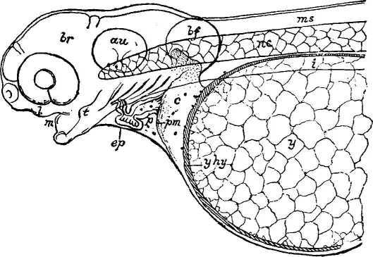
Head and Yolk Sac
EXPLANATION OF FIGURE. Head and fore part of yolk sac of young shad, just hatched, enlarged 35 times. [y], clear yolk masses involved in a protoplasmic mesh-work; [yhy], palish amber yolk hypoblast, which forms the innermost covering of the yolk; from its anterior portion blood-cells are observed to be budding off into the segmentation cavity [c]; some are also noticed in the pericardial space [p], and in the heart; [p], the posterior pericardial membrane joined to the heart and fused with the external layers [ep] a little way below [pm]; [i], intestine; [nc], notochord; [t], rudiment of tongue, seen through the transparent walls of the cheeks; [m], mouth ; [l], margin of upper lip; [br], brain; [au], ear; [bf], breast fin; [ms], spinal cord.
In order that the process may be made clear to the reader, I have represented the anterior portion of an embryo shad in the accompanying figure, in which the relation of the heart to the yolk is shown as distinctly as possible. The sketch represents the head end of the embryo with the greater part of the yolk bag within the field of view, the whole being treated as a transparent object enlarged about 35 times.
In order to understand the process of yolk absorption, to be hereafter described, it will be necessary for us to know the relation of the yolk to the rest of the embryo. The great mass of the yolk in the shad embryo at the present stage of development is composed of coarse, irregular masses of very clear protoplasmic matter.
These irregular masses in turn seem to be separated from each other by a material which occupies the fine interstices between them. It, again, is a protoplasm optically different in character from that of the
---------------------------------------------------------------------
The Bulletin of the United States Fish Commission 1882.
Page 180
masses which it envelops. The relation of the clear masses to the mesh work in which they are involved is shown in the figure. At the surface of the yolk mass, and next to the homogeneous wall [yhy] of the yolk sac proper, the clear masses become smaller, and in sections, if they may be implicitly trusted, they sometimes present the appearance of minute spheres or corpuscles. The immediate superficial envelope or covering of the yolk sac [yhy] is homogeneous, both in the living embryo viewed as a transparent object, and also when examined in carefully prepared sections. This superficial layer is different, again, in optical appearance from the clear masses and their matrix, already described, so that the organization of the yolk is found to be quite complex.
This layer also covers the whole yolk, which is, therefore, truly a closed
===============================
sac or vesicle. In color this yolk covering is palish amber, quite different from the clear body of the yolk, and at the anterior portion of the sac it is usually thicker than at any other point. In fact, just behind the heart, which is inclosed in the space [p], this superficial layer is often heaped up in the form of a conical prominence, thus becoming several times thicker at this point than at any ofher place. On the upper or dorsal aspect of the yolk vesicle there is a longitudinal depression or, furrow, along which the superficial yolk envelope is depressed. In this depression lies the cylindrical fore-gut [i], which, for the most part, afterwards becomes the oesophagus of the more advanced conditions of development. There is no connection of any kind between the intestine and the yolk sac at any time, such as has been described as connecting
------------------------------------------------------------
The Bulletin of the United States Fish Commission 1882.
the yolk and the intestine in some embryo sharks. The general form of the yolk vesicle, as may be seen in the figure, is ovoidal, slightly flattened on its up-per side, with a depression or furrow traversing the flattened portion lengthwise. It is entirely surrounded, on all sides, by a space filled with a serous fluid. At the anterior end of the yolk vesicle this space, in the stage of development here described, is most capacious, and comprises all that cavity marked by [c] between the posterior pericardial membrane [pm] and the yolk envelope [yhy]. This space [c] I have identified with the segmentation cavity, for reasons which it will not be necessary to present in detail in this place. If the heart does not actually develop within this cavity, its immediate connection with this space is an incontestable fact. Practically the heart develops within it, as we have elsewhere described. In the cod (Gadus) the mesoblast from which the heart is developed lies upon this space, and as development proceeds each step of the heart's evolution may be watched most satisfactorily.
At most, the only separation between the pericardial space [p] and the heart is effected by the development of the posterior pericardial membrane [pm], which is usually of extreme tenuity in the stage of development here
described. In fact, I am not sure that the membrane [pm] may not be perforate, for the reason that blood corpuscles are almost always found in the pericardial space at about this stage of development.
In Tylosurus I am quite sure that the pericardial cavity is not shut off from connection with the homologue of [c], because in that genus it is crowded, in some stages, with blood corpuscles, which vibrate in unison with the pulsations of the heart in the fluid in which both are immersed.
In the shad, as in other species, the membrane [pm] is continuous with the splanchnopleural or peritoneal layer. As the venous end of the heart, just above [p], pulsates, the membrane [pm] is pulled back and forth by the action of pulsation. Moreover, the membrane [pm] is continuous with and joined to the venous end of the heart, just above [p], and in front of [c]. In fact, the heart opens freely into the
cavity [c]. Free communication is thus established between the cavity of the heart itself and the segmentation cavity, or the serous space which surrounds the yolk vesicle.
We are now ready to comprehend in a measure the manner in which the material of the yolk is generally broken up into small spherules or corpuscles and sucked up out of the space [c] by the heart, and carried into the body of the embryo to be appropriated in the processes of further development. On the surface of the structureless membrane [yhy] careful observation will reveal the fact that minute spherical prominences are developed. If one will be content to observe patiently for a couple of hours, these bodies will finally be seen to free themselves from all further connection with [yhy] and to drop freely into the surrounding fluid. These corpuscles are quite colorless and present the irregular globular appearance of the white blood cells found in human blood. Owing to their continual vibration in the serous medium
------------------------------------------------------------
The Bulletin of the United States Fish Commission 1882.
in which they are found, on account of the persistent pulsation of the heart, I found it was impossible to ascertain whether they manifested any amoeboid movements or changes of form such as may be observed in the colorless blood cell. In the figure I have exaggerated, the number visible at one time at this stage in order to show more clearly the steps of the process. In the embryo shad they are formed sparingly at first, but at a later period of development they become more plentiful.
In Tylosurus they are, however, formed in such myriads at about this same stage that it would be quite impossible to count them, the serous fluid surrounding the heart being charged with vast numbers, which are in this instance, however, already reddish in color, which is not the case with the shad, where the red coloring matter of the blood, appears to be developed at a later stage. It is a singular general truth
that, in those species in which the pigmentation of the body takes place early or while the embryo is still within the egg, the blood cells become reddened much earlier than in those in which the pigmentation is delayed or retarded. In fact, it also appears to be generally true that the first lines of pigment cells are developed along the courses of the great blood vessels and in the neighborhood of specialized sensory organs.
This, however, is leading us away from the subject in hand. As the yolk vesicle or sac diminishes in bulk it tends to become pointed anteriorly. The external layer of the yolk [yhy], which, as we saw, affords the material for new supplies of blood cells, becomes thickened anteriorly, and sometimes even appears to be extended into a conical point directed towards the venous and open end of the heart. This would indicate that the yolk was being consumed from its anterior extremity.
The anterior conical end of the yolk vesicle sometimes presents a granular, or rather corpuscular, appearance after two-thirds of the whole has been absorbed. This condition is in keeping with what we observed at an earlier stage, where, as in the figure, we saw the outer layer of the yolk gradually break up into corpuscles or spherules, which were taken up by the heart, although as yet there was no evidence of a complete circulation. Together with this diminution in the volume of the yolk
vesicle, the membrane [pm] is drawn back, at its outer attached border, becoming more funnel-shaped; into this infundibuliform backward prolongation of the posterior pericardial membrane the conical anterior extremity of the yolk mass extends. Gradually the bulk of the yolk diminishes still more until it remains as a fusiform mass which is no longer prominent on the ventral side of the body of the young fish.
Meanwhile the liver of the young fish has been more developed and the portal vein makes its way over the dorsal aspect of the yolk towards the venous end of the heart. It appears probable that what now remains of the yolk may be taken up in part by the portal vein, but of this I am not well assured, further than to state that the portal vessels or channels appear in part to traverse what was formerly the segmentation
cavity [c].
------------------------------------------------------------
The Bulletin of the United States Fish Commission 1882.
The peculiar, homogeneous protoplasmic wall of the yolk vesicle [yhy] persists to the last, as I have learned from sections prepared from embryos in which the yolk sac was almost entirely absorbed. It would therefore appear that the central clear portion of the yolk [y] was by degrees transformed into the superficial palish amber layer which forms the covering of the vesicle or sac. Of the forces at work in effecting
this transformation, we know nothing more than of the efficient cause of development itself.
Thus far we have discussed the absorption of only that part of the yolk which remained after the embryo shad had left the egg. As we know that the volume of the embryo previous to hatching is greatly in excess of the volume of the germinal disk, it is fair to infer that in addition to the mode of yolk absorption here described there must be another which will account for the growth of the embryo before its heart has
developed enough to be an active agent in the process of yolk incorporation.
This second method of yolk absorption has been called intussusception, and is the primary or initial mode. It supposes that the embryo appropriates a part of the yolk during the early stages of development by a direct process of incorporation without the aid or intervention of a blood vascular apparatus, as rudimentary even as that which we have ascribed to the embryo shad. The body of the embryo, superimposed
as it is upon the yolk, is supposed to derive portions of material for further growth as these are needed from an "intermediary layer" (Van Bambeke), which probably corresponds to our palish amber yolk envelope which covers the clear yolk material in the shad.
This layer, called the couche haematogene by Vogt in his embryological history of Coregonus palaea, therefore appears to play an important part in the development of the blood at all stages both before and after the functional development of the heart. Under whatever name we know it, it is undoubted that in this layer a process of cell and blood-cell differentiation takes place. This statement is grounded on two sets of facts; namely, the observation of free nuclei in this layer by embryologists, and the undoubted circumstance of the origination of blood cells from its surface. Blood cells, especially white ones, are known to be nucleated, and no others are at first formed in the shad; it therefore follows that the nucleation must occur in the layer here understood.
Kupffer* has alluded to a similar process, but from what I have been able to gather from his writings he does not seem to have been clear in his understanding of the layer, confounding it with the true hypoblast. This opinion I was also led to adopt in my essays on the Spanish mackerel and silver gar, but I am now in doubt whether this view can be justified. My main reason being that I have been unable to discover any evidence that the intestine of the shad originates from this yolk envelope in sections prepared from such stages
Footnote * Beobachtungen uber die Eutwickelung der Knochenfische.
Arch. fur Mik. Anat., iv, 1868.
------------------------------------------------------------
The Bulletin of the United States Fish Commission 1882.
Page 184
as ought to have exhibited it. In fact, the tract from which the intestine originates is independent of this outer yolk wall from the first. The rudiment of the intestine, before the development of its internal cavity, is merely a flat band of cells somewhat thicker in the middle line than at its edges, and lies just below the tract in which the aorta and cardinal veins are afterwards excavated.
Upon referring to some of my notes, bearing the date of February 27, 1882, in regard to the structure of the yolk sac of the land-locked salmon, I find the following recorded: "As to the structure of the yolk sac, in making a dissection of a lively embryo, in a neutral salt solution, the epithelial (epiblastic) layer was found to be quite free from the yolk, so that it could be stripped entirely off from the surface of the latter." It evidently was not continuous with the subjacent layer traversed by the complex blood-vessels of the yolk, but between the two there was all exceedingly thin serous space. "On the ninth day after hatching, large numbers of red blood corpuscles were still found in the pericardiac space. Later, in diseased, or rather in what were probably injured specimens, numbers of which were kindly brought me by Mr. Fred Mather for study, I found large quantities of blood-cells in the serous space between the external or epiblastic and somatopleural covering of the sac and the vascular layer. In some cases the posterior portion of the former was abnormally much distended, so that a large cavity was developed."
To continue the reproduction of my notes, however, I further stated: "Beneath the outer layer and forming the inner wall of the serous space around the yolk, came the vascular hypo-blastic stratum in which the vitelline network of blood-vessels was developed. This, like the outermost layer, could be removed entire from the contained yolk. The segmentation cavity, with which I identify one of the serous spaces so resulting, may be either between the epiblast and vascular splanchnopleural layer, or between the latter and the yolk." But the homologue of the segmentation cavity is probably the latter. Inside of the vascular layer I encountered the yolk vesicle proper, comparable with the palish amber layer of the shad. In its superficial portion I find the oil spheres immersed. This stratum in fact is the "couche haematogene" of Vogt, which is as well developed in the embryos of Coregonus albus of our lakes as in the European species, studied by the versatile naturalist of Geneva.
Here as in many other species there is a tendency of the blood channels to present the appearance of irregular wide passages over the yolk, somewhat lacunar in nature. This feature is observed, however, only in such as have a vitelline circulation, as, for example, in embryos of Apeltes, Tylosurus, Carassius, Idus, Fundulus, Esox, Goarces, Salmo, etc., and not in Alosa, Cybium, Parephippus, Pomolobus, Gadus, which are without a vitelline vascular system. But these two types run into each other, for in some the intestinal or portal or else the median subintestinal system of vessels may hereafter be found to take a share in the process of blood development.
------------------------------------------------------------
The Bulletin of the United States Fish Commission 1882.
I have elsewhere* alluded to the researches of Gensch,** who investigated the development of the blood of Zoarces and Esox. He observes that the blood originates in these forms by budding from the hypoblast, and credits Kupffer with having been the first to call attention to the fact. I cannot help thinking, however, that what he means by the hypoblast is really the equivalent of the palish amber envelope of the yolk of the shad, and in no sense anything but a temporary and evanescent structure, which vanishes completely when the contained yolk material has been absorbed. It may be proper, perhaps, to designate this structure by the name of yolk hypoblast, but beyond the name it is doubtful whether it is proper to imply more, because I have yet to learn, after careful investigation, that it ever enters into the formation of any of the organs or membranes of the body cavity in which it is actually inclosed.
Before concluding, however, I wish to call attention to one more difference between the embryo of fishes with a vitelline circulation and those without it. In those forms in which the blood-vascular network covering the yolk is well developed the hypo-blastic vascular layer is relatively thick and distinct in cross-sections. In those in which there is no vitelline circulation the reverse is the case.
When we come to examine cross-sections, the epiblast, mesoblast, and the true hypoblast are so intimately united and their combined thickness so slight that it is with great difficulty that they are resolved with the microscope. In the young shad, directly after hatching, the outer covering of the yolk is extremely thin, and measures about one two-thousandths of an inch in thickness. Immediately beneath it and separate from it lies the homogeneous wall of the yolk vesicle.
This structure, which we have chosen to call the yolk hypoblast above, is, on the contrary, often ten times as thick as the outer and external yolk envelope which comprises, as we saw, all of the embryonic layers, but which have been reduced to the greatest tenuity.
From the foregoing recital of facts we are led to a somewhat clearer understanding of the method of yolk absorption as observed in young fishes. We cannot help admiring the simplicity and efficiency of the apparatus. Whether the space identified by me as the segmentation cavity in fishes must be considered equivalent to the pleuroperitoneal space in the embryos of birds, I am unable to state; this is, however, probable. Practically, there is very little difference between the mode of yolk absorption, as manifested in the chick and in the fish. If by a large license, as it seems to me, we admit that the yolk vesicle of the shad is really its hypoblast, the origin of the blood and the incorporation of the yolk substance are similar in birds and fishes. In the latter the nature of the hypoblast may be so obscured that I may have fallen into error in not regarding the yolk wall as hypoblastic; however, that
Footnotes
------------------------------------------------------------
The Bulletin of the United States Fish Commission 1882.
a structure which disappears so entirely as not to leave behind any organ which may be with certainty traced to it as its source of formation, I am loath to regard as one of the primary embryonic layers. The yolk is entirely included within the abdominal cavity in fishes as soon as the blastoderm has closed over it. In this regard it widely differs from the chick, a point to be borne in mind in this discussion. The serous space around the yolk in the shad represents the body cavity. Looking again, during the present writing, at sections made from embryos shortly after the inclusion of the yolk by the blastoderm, I am convinced more forcibly than ever of the correctness of the view herein maintained.
I cannot persuade myself, even while examining this early stage, that
the yet thin and incipient yolk wall is continuous, or likely to have been
with any of the embryonic layers, except during the very earliest stages
of development and before the differentiation of the layers. The yolk
hypoblast, therefore, has only a physiological and mechanical function to
perform, which ends with the final and complete absorption of the yolk
out of the serous abdominal cavity.
Immediately after the heart is formed, as soon as it begins to pulsate,
and long before hatching, it seems to open directly into the serous cavity
already described. In this condition and even much later it seems
to the observer almost like an independent being within the embryo,
sucking up the yolk; an appearance which, at this time, is of course
illusive, as the breaking down of the yolk by the help of germination
and the circulation probably does not begin until about the time the
embryo is free from the egg.
Previous to that time the appropriation of the yolk material probably goes on by intussusception, as already mentioned. The communication of the heart with the serous cavity surrounding the yolk, as stated before, is direct, but as soon as the Cuvierian ducts are developed, its venous end is almost entirely fed by them
from the cardinal veins, the serous cavity in front of the yolk only communicating imperfectly with the heart. In four or five days the bulk of the yolk is absorbed, some remnants of it sometimes remaining for a long time afterwards, or up to the tenth day or even later. The rate of yolk absorption is profoundly influenced by temperature, which is no more than was to be expected.
The diminution in the bulk of the yolk is accompanied by a gradual collapse of the outer sac, the diminution of the capacity of one seeming to keep pace with that of the other. This is the case with the shad, and in fact with most embryo fishes. The most notable exception to this rule being the very remarkable phenomena first observed by the writer in the embryos of Cybium and Parephippus, where the collapse of the yolk mass in its vesicle, as absorption goes on, is not followed by an immediate and equivalent diminution of the capacity of the external sac. It follows from this state of affairs, in these species, that the serous cavity around the yolk becomes remarkably enlarged. The question, then, also arises, how does the extra water find its way into this
------------------------------------------------------------
The Bulletin of the United States Fish Commission 1882.
serous space, unless by a purely physical process of transudation, or
osmosis of water from without, which keeps pace with the collapse of
the yolk, the absorbed water taking the place of the latter as it
diminishes in volume?
II-NOTICE OF AN EXTRAORDINARY HYBRID BETWEEN THE SHAD AND STRIPED BASS.
A number of young fish which had already lost their yolk sacs, in consequence of which it is to be supposed that they were already several days old, were received from Havre de Grace at the central station on the evening of June 13. They were immediately placed in an aquarium, but mauy of them died in a day or two after, save about fifty, which were transferred by the writer to one of the smaller of the carp
ponds in charge of Dr. Rudolph Hessel, where, as Professor Baird had suggested, they might possibly find some food suited to their wants and grow large enough for us to learn something of their future history.
The case is an extraordinary one, as the possibility of interbreeding members of such very distinct families as that of the Clupeoids and Percoids, unless the impregnation took place under the very eyes of the naturalist, might well be doubted, as even such a thing as the successful impregnation of the ova would naturally be doubted by those familiar with the recorded facts related of hybrids in general. The evidence in favor of the fact in this case is, however, too strong to be passed over, and until we know more of the later history of this singular hybrid, the following notes on the differences which were presented by the embryos as compared with those of the true shad must suffice. The striped bass was the male and the shad the female parent.
Teeth more numerous and more hooked on the lower jaw; at least three pairs, only two pairs in shad of same age. Lower jaw itself longer, with gape of mouth much wider; ear capsule proportionally much larger than in shad larvae of same age, and otoliths much larger. Tail a little more fan-shaped than in shad of same age, and pigment and fine cellular radii of fins slightly more developed than in the latter. Intestine much more slender, that is, its lumen is much less spacious than in Alosa or
Clupea. Liver in about the same position as in larval Alosa, but gall-bladder and eye relatively and perceptibly larger; Meckel's cartilage a fourth longer. General form that of the larval Alosa, but head more prolonged and acuminate anteriorly. The preponderance of characters appears to be towards the female parent, and appears to be an undoubted hybrid.
The eggs were taken by some of the crew of the steamer Fishhawk, at Havre de Grace, and were impregnated with the milt of the "rock" or striped bass, because no ripe shad milters happened to be at hand.
------------------------------------------------------------
The Bulletin of the United States Fish Commission 1882.
III. CAUSE OF THE NON-DEVELOPMENT OF FUNGUS ON THE EGGS
HATCHED IN THE MCDONALD JAR.
The development of fungus on shad eggs, as far as we are able to judge, has always been due to the conditions under which they were placed. When any imperfection existed in the current of water flowing through the cones, the eggs which would collect on some spot on the bottom screen which had been partially choked up with sediment, caused both dead and live eggs to collect in a mass over such places.
The fungus, on account of its very rapid development, when once started amongst such lots of eggs, would soon mat them together in large masses, which had to be removed with the small "skim net". The absence of any current amongst masses of ova seems to be the one favoring condition under which the egg-fungus grows most advantageously. The mycelium, once established on the membranes of a lot of eggs, soon attacks those which some movement may bring into contiguity with those already infested.
The plant possesses all the features of a parasite converting the material of the egg into its own substance. Its reproductive activities are also developed very early, and its germs are produced in vast numbers, which are very minute motile bodies which escape from their receptacles on the parent plant to pollute the surrounding water. It is easy on this account to understand that any apparatus from which it is impossible to effectually remove dead eggs, and in which there is an imperfect circulation of water amongst the latter, would favor the development of fungus and the destruction of many ova.
Formerly the Bell and Mather cone was disposed, if not carefully watched, to favor the development of fungus. Recently this objectionable feature seems to have been overcome to a certain extent. No "cone" yet devised is, however, as good as any one of three different forms of glass apparatus; the Chase, the Clark, or the McDonald jar offer advantages over any form of metal apparatus. These systems of glass-hatching vessels can be kept so thoroughly free from dead eggs without a skim net, and the circulation can be so perfectly regulated so as to keep every egg in continuous movement, thus preventing any fungus spores from lodging on the eggs.
The continuous and gentle attrition of the ova in the glass jars effectually prevents any fungoid germs from adhering to the membranes of the ova; the pest, which is in this way prevented from obtaining a foothold, never causes any serious trouble.
Another advantage offered by the glass jars is the ease and accuracy with which the number of eggs may be estimated by graduating the jar into inches or into intervals indicating the spaces occupied by single thousands of eggs, or by measuring the height of the column of eggs in the jar with a graduated rule indicating similar quantities. This also enables the person in charge to estimate very closely the number of dead eggs which accumulate on the surface of the live ones in a layer of nearly even thickness. This is impossible in the metal cones, and the estimate of losses has hitherto been little better than guesswork.
------------------------------------------------------------
The Bulletin of the United States Fish Commission 1882.
In the glass vessels the estimate is very nearly accurate and very easily made.
In hatching white perch or other adhesive eggs, if the strings with ova adhering to them were hung into the McDonald jar, into which a quantity of shad ova had also been introduced, I think it altogether probable that the attrition of the shad eggs against the perch eggs would prevent the latter from becoming infested. The shad ova in their rolling movements over the others would tend to prevent the lodgment of fungus spores, as already pointed out in my discussion of shad ova.
In the English edition of Maout and Decaisne's Botany, p. 975, I find the following account of the egg fungus or alga as it is indifferently called by different authorities. In order to disseminate a fuller knowledge of its life history I will here reproduce what these distinguished writers say of it:
"These singular vegetables are considered to be fungi by some botanists; they live, in fact, on organic matters in a state of decomposition in water, where they act upon oxide of iron by decomposing the carbonic acid, absorbing the oxygen, and thus setting free the sulfuretted hydrogen, which destroys the vegetables or animals near it. [This indicates the great importance of at once removing from the hatching vessels any masses of fish ova which have become infested.] Notwithstanding the significance of these biological phenomena, several physiologists who have carefully studied Saprolegnieae do not hesitate to class them amongst Algae.
'Saprolegnia ferax', says Thuret, 'is usually found on the bodies of drowned animals, which it covers with a whitish down; it even attacks live fish. Nothing is easier than to procure this singular Alga. Let a vase be filled with water from a garden tub, and some flies be thrown into it, and it will usually be developed in a few days. The body of the fly becomes covered with hyaline filaments, which radiate around it, enveloping it with a whitish zone. Under a microscope, these filaments are seen to be continuous, simple, or scarcely branched, and to contain minute granules, which show a motion resembling that which is seen in the hairs of Phaenogams.
These granules are very numerous, especially towards the upper extremity of the tube, to which they give a gray, somewhat russet tint. This portion soon becomes isolated from the rest of the filament by the formation of a diaphragm. Then the contained matter coagulates in small masses, which become more and more sharply defined, and end by forming so many zoospores. These phenomena succeed each other very rapidly; often in less than an hour the granular matter becomes condensed at the top of the filament, the septum forms, and the zoospores appear. Finally the tube, which has a small protuberance at its extremity, bursts there, and the zoospores escape, the first with impetuosity, the others more slowly; they are turbinate in shape, and furnished with two hairs. This is not the only mode of reproduction possessed by Saprolegnia; another phenomenon succeeds. The filaments emit small
The Bulletin of the United States Fish Commission 1882.
lateral branches, the extremities of which swell into sacs of a blackish hue, due to the condensation of their granular contents. Soon a septum forms, isolating the sacs from the little tubes which serve as pedicels to them. After some time the granular matter divides into several masses, which at first adhere to the walls of the sac, but which later become free and spherical. Sometimes there is only one of these masses; sometimes the same sac contains fifteen or twenty. I have fancied that I could recognize on their periphery little mammillae resembling regularly arranged opercula."
The sacs have been termed by Pringsheim oosporangia. The oosporangia require fertilization to enable them to produce fertile spores. It is obvious, therefore, that Saprolagnieae have a double mode of reproduction, similar to that of Vaucheria; the one asexual by means of zoospores; the other sexual, producing oogonia arising from the fertilization of a sporangium (oosporangium)."
The tubes alluded to in the first portion of this quotation, when developed on the surface of a dead shad-egg, stand out in all directions like a vast number of rays; to make a vivid comparison, the infested egg looks very like a seed-head of the common dandelion, with all of its slight, tufted seeds still adherent to the receptacle. The zoospores alluded to as possessing two hairs or filaments have these latter endowed with a power of movement; these filaments in turn propel the body of the spore about, so that in this way the noxious germs of the plants are widely distributed through the water.
IV. EXPERIMENTS WITH CARBOLIC ACID TO KILL THE FUNGUS ON LARGE FISHES.
Several hybrid gold-fish in the aquaria in the central station became badly infested with fungus, probably because too large a quantity of dead shad-eggs was thrown into the water to serve for their food, which, instead of being immediately consumed, remained lying on the bottom of the tank until the fruiting condition of the egg fungus was developed on them. These spores from the egg fungus then lodged upon the skin of the fishes and commenced to grow, showing the mode in which the fungoid infection might be conveyed from the eggs to the adult fish.
Knowing the fungicidal properties of carbolic acid, it occurred to me to try a very weak solution in water to see if it would kill the fungus on the fish. A badly infested fish was placed in a glass aquarium holding nearly four gallons of water; in the first trial ten drops of a concentrated No. 4 solution of carbolic acid was dropped into the water, with little more effect than to make the fish exceedingly restless. The next trial was made by doubling the quantity of acid used, which in the course of ten minutes showed that the fish was suffering and would probably die if fresh water was not immediately turned into the aquarium to replace that in which the acid had been dissolved. In a day or so afterwards the fish experimented upon died from the inroads of
------------------------------------------------------------
The Bulletin of the United States Fish Commission 1882.
the fungus, showing apparently that the acid was not the cause of its death.
Mr. Livingston Stone has recommended the use of a strong solution of common salt to kill fungus, which he has found quite effectual. Mr. Behler, of the Druid Hill hatching house, says a saturated solution of salt water is effectual; and he also states that if young salmon infested with the fungus are dipped bodily into asphalt, the fungus will be killed and the young fish come out all right and healthy, the asphalt gradually peeling off of their bodies. This last statement seems almost incredible, but it has been stated to me in good faith by the experimenter himself.
4. DISTURBANCE OF THE BALANCE OF CONDITIONS, AND ITS INFLUENCE ON THE CRUSTACEAN FOOD OF THE SHAD.
In conducting the experiment of feeding the young shad, vast numbers of minute Daphnidae were caught, which were put in the glass aquaria with the young fish; sometimes the number of these minute crustaceans captured at one time was so great that when closely packed they would almost equal a solid quart in bulk. Great mortality was noticed among them soon after being placed in the aquarium with the fish, which would indicate that they had been subjected to a fatal change of surroundings.
Various reasons might be assigned as probable causes of this mortality. It may be that the rapid circulation of water in the aquarium was one of them, or it may be supposed that when taken from the stagnant water of the carp ponds and transferred to water of a considerably lower temperature in the hatching house the change was too great. A still more probable cause may be the fact that the crustaceans, when removed from amongst the water plants in the pond, were deprived of their natural food, and as a result starved to death.
Whatever may have been the cause of this phenomenon, however, we may be assured of this fact, that in supplying live food to shad larvae we must also be careful to attend to the vital conditions required by the former. If it is desired to keep this living food in a healthy state, so as to multiply in the aquaria, it is probable that water plants must be supplied for the purpose of furnishing the requisite conditions for the protection and multiplication of the food of the living shad-food. Until recently we have not been able to supply quite the proper conditions for the nutrition of the young shad, and in feeding the latter it becomes evident that we must take care to feed the food, which may be done by providing the conditions for the propagation of protozoa, algae, etc., in the aquaria or shad nurseries.
VI. A MEANS OF DEMONSTRATING CARTILAGE IN FISH EMBRYOS.
Knowing the potency of potassic hydrate in dissolving protoplasm, it occurred to me to try its efficiency as a means of getting rid of the tissues and membranes which envelop the trabeculae cranii, hyoid, and
------------------------------------------------------------
The Bulletin of the United States Fish Commission 1882.
branchial cartilages of the shad. A 5 per cent solution it was found would rapidly dissolve the dermal, neural, and muscular tissues, leaving only the rudimentary, aponeurotic membranes and matrices of fasciae between the muscular segments and the notochordal sheath. The head cartilages remained undestroyed and could afterwards be stained. The method is useful, however, only where fresh material is at hand, as it is almost impossible to mount a specimen satisfactorily for permanent preparation. In staining, it is desirable to wash out the alkali as much as possible, and afterwards to investigate the arrangement of the cartilages of the skeleton under gentle pressure under a Fol's compressor.
VII. METHODS OF HANDLING WHITE PERCH OVA.
The egg of the white perch is notoriously adhesive, and for this reason is one of the most troublesome to deal with practically. The eggs were taken upon cotton yarn, which was drawn up through a funnel, into which the eggs and milt had been squeezed from the spawning fish. The cord, covered with the adhering eggs, was then wrapped upon a wooden reel and sent under cover of damp cloths to the central station, where they arrived in fine condition, almost every egg being impregnated. This system, devised and carried out under the superintendence of Colonel McDonald, was really another adaptation of the dry method of carrying the ova of the shad.
After reaching the central station, the cotton cord, with the adhering eggs, was cut into lengths of 10 to 12 inches and suspended in the glass hatching jars. The development progressed normally as long as not interfered with by the growth of saprolegnious fungus. There being no attrition between eggs, as amongst shad ova, when incubated in the jar, fungus soon established itself, and grew until the whole brood was practically destroyed.
Another mode was to introduce the wooden reel, with the eggs adhering to the cord, into a wide aquarium. These also were attacked by fungus, but slightly more favorable results were obtained. With the water at 58� F. to 60� F., the ova, hatched out in six days. The water in the jars, for some of the time, stood at 51� F to 55� F and rose but little above this point.
The use of narrow strips of glass or mica might, it seems to the writer, be used to advantage, or even glass plates would be convenient, upon which the eggs might be allowed to adhere. It was discovered this spring by the writer that where the eggs were allowed to stick to the string in several layers, the uppermost strata seemed to smother or prevent the respiration of the lowermost layers, which were killed in consequence. This is opposed to the experience of Mr. Clark with the Atlantic herring, and also to that of Capt. Z. L. Tanner with the Branch herring. Both of these experimenters found that great masses of the eggs would hatch with scarcely any loss, although it was scarcely possible to understand how it was possible that there should be any circulation of fresh water through such large clumps of adherent ova.
------------------------------------------------------------
The Bulletin of the United States Fish Commission 1882.
The nature and origin of the material by which adherent ova are made to stick fast to foreign bodies and to each other is an interesting inquiry. It appears to be a mucous substance, derived, possibly, as a peculiar secretion either from the ovarian follicles or some special glandular structures within the ovary. Its remarkable property of hardening under water I find to be characteristic of the material in all the species with adhesive ova which I have yet had the opportunity of examining.
VIII. NOTES ON SMALL FISHES AND WATER ANIMALS WHICH PREY ON FISH LARVAE.
Recently (June 1882) some four-spined sticklebacks (Apeltes quadracus)
in spawning condition were received at the Central or Armory hatching station of the United States Fish Commission from Mr. W. P. Seal, of Philadelphia, who, in his letter announcing their shipment, informed me that they would not accept dead food, but must be supplied with small living insect or crustacean prey, which they would themselves capture. The specimens were mostly males, and would measure an inch and three-eighths in length; some were smaller.
Mr. Seal's opinion as to their feeding habits was soon verified, as it was found that dead food had not the slightest attraction for them, but as soon as live food was offered them they exhibited a vivacity and alertness in its capture which was truly surprising. I obtained a supply of this from the government carp ponds by skimming the surface of the water amongst a rank growth of aquatic plants with a fine net, where it was found that Daphiniidae, neuropterous, and coleopterous larvae abounded, as well as numerous brown aphids, which had blown on to the surface of the water from the taller plants fringing the pond.
These creatures were transferred to the aquarium containing the sticklebacks, when the work of destruction was at once commenced. The lively little ichthian marauders would poise themselves in the water, roll their eyes, and when one of their victims was within sure range they would pounce upon it, rarely missing their mark, in spite of the fact that the latter, as in the case of the Daphnids, might happen to be quite minute. Hard-shelled coleoptera and bugs were not accepted, nor did the larger neuroptera seem to attract their serious attention. The smaller, softer-bodied animals seemed to be most palatable.
Upon observing this, it occurred to me to try them upon shad larvae. About twenty-five of these, three days old, were then put into the jar; no sooner than they had made themselves conspicuous by their active wriggling movements through the water, the keen eyes of the sticklebacks perceived them; their destruction was completed in about half an hour by the half-dozen individuals of Apeltes. The experiment was repeated with a similar result. showing the destructive capacities of Apeltes when brought to bear upon helpless shad larvae. Gunther has noticed the voracity of Gasterosteus in his "Study of Fishes."
It has been maintained by some writers that fish larve, which in the
------------------------------------------------------------
The Bulletin of the United States Fish Commission 1882.
case of many species are very transparent, are protected by this transparency, which renders them to a great extent invisible in water. The transparency of the larval shad is notorious, and with the exception of the glittering iris and two dark lines of pigment cells above and below the intestine, would be almost invisible in the water; yet it is evident from our experience that this transparency affords little or no protection from so sharp-sighted an enemy as the stickleback.
And if it could be shown that one stickleback under natural conditions could devour a dozen shad larvae in an hour in the weedy flats of rivers, where the naturally spawned young of Alosa may be supposed to abound, the destruction of such larvae during a season must be enormous in respect of numbers. The foregoing observations tend to show, it would appear to the writer, that the transparency of larval fishes is practically of no avail in the presence of the predaceous species of their own class.
If shad larvae are visible to sticklebacks, there is no valid reason why they should not be equally so to young predaceous fishes of a dozen other species, and if, as we may suppose with good reason, not unsupported by observation, that young fishes will be attracted to weedy flats on account of the insect prey which there abounds, we may be almost equally certain that any very young fish larvae, although still transparent, will not escape the vigilant eyes of their finny enemies, and consequent destruction.
The larvae of many neuropterous insects which undergo their transformations in water, as well as those of certain coleoptera, such as Dytiscus, are noted for their predaceous habits. I have seen the larvae of the dragonfly capture and devour a young salamander almost or quite as large as itself. These gourmands of both the neuropterous and coleopterous order are probably not excelled in rapacity by any small fishes, though their rapidity of movement is probably not as great as that of the latter. Now as to the facts of the case, I find upon trial that both neuropterous and coleopterous larvae are capable of destroying young shad, but to what extent I am not assured, as a sufficient number of observations are wanting.
The main point at which we have been aiming is, however, clear. Transparency is no safeguard from either active vertebrate or invertebrate enemies, and now the question arises as to the point already urged in a former paper by the writer, as to the expediency of setting shad larvae free in weedy shallows in rivers where it is found that their natural food abounds, together with their enemies. The only answer that seems possible under the conditions as we now know them is the following: That it is best to put the larvae where they will soonest find food, although they be at the same time brought into the presence of the greatest number of enemies. The chances of survival seem to me to be greater where the food is most abundant, for the following reason: Notwithstanding the fact that many enemies may be present, the chances to obtain food in such places are so much more favorable, so that the growth and
------------------------------------------------------------
The Bulletin of the United States Fish Commission 1882.
vigor of the larvae will be soonest enhanced, thus enabling them to grow faster, become more vigorous, and the more readily able to escape their enemies.
IX. OBSERVATIONS ON THE FOOD OF THE YOUNG JAPANESE GOLDFISHES.
Professor Baird, noticing that the young fish in the carp ponds appeared to be feeding very actively upon something, requested me to investigate the contents of their stomachs and intestines. On the 6th of June I opened a specimen 30 millimeters long, in the intestine of which I found the following:
Dirt and particles of quartz-sand.
In the stomach of the same specimen I found the following:
In another specimen, besides the foregoing, I found the following:
------------------------------------------------------------
The Bulletin of the United States Fish Commission 1882.
In still another specimen I found both the dermal and flesh spicules of a fresh-water sponge and the remains of a portion of a tracheal tube of an insect.
A more exhaustive examination of the fish, which I did not care to sacrifice to any great extent, would have shown that in feeding they had probably laid the entire fauna and flora of the pond under contribution; at any rate, the foregoing list shows that there was no want of variety in the make-up of the bill of fare consumed by these young Cyprinoids.
X. EXPERIMENTS IN SUPPLYING THE PROPER FOOD FOR LARVAL SHAD.
It was suggested to me by Professor Baird in 1880 to try to discover the kind of food upon which the young shad normally feeds, and if possible to collect or breed the food in quantities large enough to afford a supply of nutriment for the larvae. My investigations upon the intestinal contents of the adults during that season taught me approximately what I might expect to find to be the food of the young. Investigations which I conducted in the latter part of the season of 1880, upon the young which had been kept for fourteen days at the navy yard at Washington by Mr. F. N. Clark, showed that my surmises had been correct.
As already discussed in my paper, published in this Bulletin, entitled "On the Protozoa and Protophytes as the Primitive Source of the Food of Fishes," the young shad, under favorable conditions, soon prey upon other minute organisms. The mouth is not open widely enough immediately after hatching to take food, nor can the mouth be opened and closed at this time, but by the time the yolk sac is fairly absorbed, or in four to five days after hatching, the young fish will already take food, as observed by us this season in the aquaria at the central station. At first the mouth is on the under side of the head, and it is only after the jaws and gill-arches have grown longer that the mouth is widely enough open to seize food with the four conical, hooked teeth with which the lower jaw is armed; this condition of development is attained when the yolk sac has been mostly absorbed.
It was a problem with me for some time as to how the food of the young shad, which consists mostly of Copepoda, Ostracoda, etc., was to be got in sufficient quantity to make artificial feeding a success. Knowing the favorable conditions which existed in the carp ponds in Washington for the multiplication of these crustaceans, I took a fine net to the ponds and found a locality where I could skim them out from water, amongst dense growths of Anacharis, by the many thousands. The skimming was continued until a large number of Copepoda and Daphniidae were obtained, which were turned out amongst the young fish into the collectors used by Colonel McDonald in connection with his hatching jar. The young shad in the collector aquarium, having vast numbers of their favorite food all about them, which could not escape
------------------------------------------------------------
The Bulletin of the United States Fish Commission 1882.
on account of a fine screen which was placed over the outlet from the collector, soon began to feed. In a half hour afterwards, I should think fully ten per cent of the young fish which had already lost their sacs had begun to feed. The evidence of this was the presence of their crustacean food in the intestine, in which it could be readily seen with the naked eye through the transparent walls of the abdomen. It would tend to accumulate just behind the origin of the liver and air-bladder, which marks the origin of the larval stomach.
These experiments were instituted on the 11th day of June, and daily supplies of Copepoda were afterwards sent to the Armory by Dr. Hessel from the carp ponds, where the latter gentleman also found other localities where these crustaceans were still more abundant than those originally visited by me. The mortality amongst the young fish, even in the presence of their natural food, was very great--not less than 75 per cent; but it was shown that it would be practicable to furnish the required food; and as I hear since I left Washington, from Colonel McDonald, the young fish continue to feed, growing rapidly and giving every promise of surviving until an advanced condition of growth is reached. Some of these young shad, according to my last advices from the central station, must now, July 6, be nearly a month old.
Since the above was written some time has elapsed, and the survivors of the lot of young shad which gave such promise of continuing to grow have either been captured for preservation in alcohol or have escaped from the aquarium in which they were confined. The last surviving specimen of the lot lived to be forty-two days old, when, it accidentally escaped into the sewer-pipe before it was possible to recover it.
Those in charge at the time inform me that this individual was about one and a quarter inches in length at the time of its escape. This would be about the length it would have reached at the age mentioned above, judging from some specimens which were submitted to me for examination from North Carolina; these having had the good fortune to survive to the age of three weeks, when they measured 22 millimeters long, or about seven-eighths of an inch. The Armory specimens, I am told by Mr. J. E. Brown, fed quite ravenously upon the living Copepoda, which were supplied to them to the last.
XI.MECHANICAL CONDITIONS AFFECTING THE DEVELOPMENT OF FISH OVA.
There is a class of facts met with in embryological observations which have a significance which, I think, have not received the attention they deserve. They relate more especially to what may be termed the mechanism or construction of ova, and to the peculiarities of development which grow out of conditions of construction as necessary results thereof.
Holoblastic ova, for instance, are very differently conditioned from the mesoblastic. Practically, we can scarcely say of the former type that it ever develops a blastoderm, but rather that the whole of it at
--------------------------------------------------------------
The Bulletin of the United States Fish Commission 1882.
once becomes blastodermic in morphological character. Nor do holoblastic ova even behave similarly to each other. Even in the progress of development the gastrula stage is sometimes so modified or interfered with by the mechanical accidents of the construction of the ovum as to be almost wholly obscured or suppressed. Such occurs in a few cases like Eucope and Stephanomia, where a gastric cavity is formed without gastrulation before the oral opening is broken through, so that the fact of gastrulation is not universally a preliminary to the formation of the archenteric cavity; formerly, in its positive form, an embryological doctrine which observed facts have shown to be untenable when universally applied.
Again, it is certain that the blastopore which results from the closure of the outermost layers of the ovum over the innermost ones, or over the yolk, does not always correspond either to stomodeum or proctodeum, or to mouth or vent, but that it is sometimes an evanescent structure of little or no morphological significance. This is the case with what is probably the true blastopore of the Teleostean ovum.
The mechanical relation of germ disk or blastoderm to the yolk in the Teleostean ovum is a peculiar one and has not hitherto received the attention which its importance has deserved. The peculiar mode of development which has grown out of this relation is in the highest degree interesting as compared with other forms. After the disk has been formed by the amoeboid aggregation of the germinal protoplasm, a series of phenomena present themselves during segmentation which will well repay attention. As soon as the segmentation is fairly under way, so that the rudiment of the embryo's axis begins to be apparent at the edge of the blastoderm, it is found that a shallow, crescent-shaped cavity has appeared underneath the central portion of the disk not immediately embraced by the embryo.
This space extends beneath the blastoderm and grows laterally with the growth of the disk. It is, in fact, a space filled with a film of fluid over which the disk may spread without friction on the underlying yolk. It remains until the blastoderm finally closes over the yolk entirely, when it may still be seen as a space, in some species separating the membranes of the embryo all round from the true yolk sac within. This arrangement, of course, causes the development to present some peculiar features, since the development of the embryo is perfectly sessile on the yolk.
Later on, the heart communicates directly with this cavity, as in Alosa and Pomolobus, and yolk corpuscles are budded off directly into it and sucked up by the heart, to be carried directly into the vascular system. Nor is this the only feature of novelty; at the tail, the rim of the blastoderm closes and forms what I have denominated the caudal plate, which afterwards enters into the development of the tail. The whole of the blastodermic rim (ringwoulst) is thus absorbed, and no similarity remains to make the Teleostean embryo comparable in this respect with the Elasmobranch, in which the rim of the blastoderm is not incorporated in this manner. In the region of the caudal plate, a solid neural cord or rod is continuous with the notochord and rudiment
------------------------------------------------------------
The Bulletin of the United States Fish Commission 1882.
of the hind-gut below, so that the gastrula is practically but obscurely realized by means of a strand of cells which connects the neurula and hind-gut. From Balfour's account I infer that the development of Lepidosteus is essentially similar. The hind-gut develops from behind forwards, and almost immediately after the tail commences to bud out the vent appears beneath the latter. The gut has absolutely no connection in any part with the yolk-sac, and the liver appears as an enteric thickening at first, and afterwards as a ventral diverticulum of the intestine, lying upon the upper and hinder aspect of the yolk. The air-bladder is a dorsal outgrowth of the intestine, which originates a little way behind the origin of the liver, from what may be termed the Anterior duodenal region. The heart develops as a simple tube in the pericardiac region, and almost from the first communicates with the segmentation cavity.
The body up to the time of the closure of the blastoderm grows, from behind forwards from the edge of the latter, adding somite to somite behind with the progress of development. In some cases the rim of the blastoderm commences to segment into muscular somites even before the closure of the blastoderm.
Comparing this form of development with that of the Amphibian and Marsipobranch, we find that we are forced to look upon the blastoderm of the Teleost, exclusive to the yolk, as their complete morphological equivalent, and that the Marsipobranch justifies the comparison in the peculiar way in which the body of the latter grows from behind forwards out of the caudal mass, just as the embryo Teleost grows from behind forwards from the edge of the blastoderm; so that, although in other respects there are some essential differences, such as in the relation and homologies of the neurula, hind-gut, and the blastopore, otherwise there is an evident similarity, which may give us a key to the comprehension of why it is that the embryo Teleost develops at and from the edge of the blastoderm.
It was not my intention, however, to enter into an embryological discussion in this place so much as to show that the development of the teleost was peculiarly conditioned mechanically in consequence of the peculiar organization of the egg, and that that had much to do in determining its mode of development. The real cause of some of its singularities appears to be the presence of the relatively enormous yolk which must be included by the blastoderm in order to be absorbed. The process of yolk-absorption itself in the teleost offers some strong contrasts with that described by writers on the absorption of the yolk of birds and Elasmobranchs, as we shall see in another place.
A few words more on the mechanical conditions presented by the ova of several genera of fishes, and I have done with this part of the subject. I am familiar with the ova of four genera of salmonoids; Salmo, Salvelinus, Coregonus, and Osmerus; all are characterized by an abundance of oil drops embedded in the vitellus, but most abundantly just immediately under the germinal disk, where indeed most of
------------------------------------------------------------
The Bulletin of the United States Fish Commission 1882.
the oil seems aggregated in the species studied by me. This oil behaves like oils in general, even in the live eggs, i.e., its specific gravity is less than water, in consequence of which the aggregation of oil drops underneath the disk tend to keep the latter directed constantly upward. If the egg is turned, the buoyancy of the oil drops at once turns the vitellus within the egg membrane, and brings it to rights with the germinal disk looking upwards. This contrivance, if one may so call it, for righting the egg is perfectly automatic. Later, when the embryos are hatched, the presence of the oil drops in the upper part of the yolk-sack helps to keep them in the natural position.
Another totally different type of eggs is that of the cusk, crab-eater, Spanish mackerel, and moon-fish. In all of these forms the egg is buoyant in consequence of the presence of a single large oil sphere. This oil sphere is always situated at a point in the egg almost exactly opposite the germinal disk. In consequence of this arrangement the germinal disk is constantly inverted; that is, it is carried on the lowermost face of the vitellus, the whole of the latter lying above it. It will be seen that in this case the buoyant oil drop of the egg acts in a manner just the reverse of what we noted in the eggs of the salmonoids.
Even after the young have escaped from the egg membrane they are at first unable to right themselves, but swim for a time upside down. This is due to the presence of the oil drop in the yolk-sack to the ventral wall of which it is permanently fixed.
The egg of the cod, strange to say, is wholly without the oil drop, but the specific gravity of the vitellus is so slight that it behaves precisely like the foregoing, and has the germinal disk constantly directed downwards, floating in this position.
The egg of Morone americana, or white perch, is another special case. Here the egg is adhesive and fixed, and embedded in the vitellus there is a very large oil sphere. In consequence of the fixed character of the egg membrane, the oil-drop controls the position of the vitellus and keeps the disk inverted and on the lower side of the vitelline globe, while the free, uncovered portion of the latter is always directed upwards, at least during the early stages.
The egg of the shad is another special case, and here there is an unusually large water space all around the vitellus between the latter and the egg membrane, but the egg is non-adhesive, and its specific gravity is greater than that of the water in which it is immersed. The peculiarity about the behavior of the egg is the constant disposition of the germinal disk to arrange itself at the side of the vitellus when viewed from above, though there is no oil whatever present in the vitellus to influence the position of the vitellus or germ.
Again, the egg of the plectognath, Aleuteres, is green, with a cluster of oil droplets embedded in its yolk or dentoplasm at one side. Its germinal matter or protoplasm is relatively large in amount.
In Fundulus and Syngnathus the oil drops appear uniformly distributed
------------------------------------------------------------
The Bulletin of the United States Fish Commission 1882.
and embedded in the superficial portions of the yolk nest to its external surface. This brings the deposits of oily matter in close proximity to the vessels traversing the vitellus. The function of these oils, aside from their buoyant tendencies, as in these last cases, is not clear, and, beyond the fact that they are evidently absorbed together with the remainder of the yolk, we know little of their nutritive properties. Perhaps, in the process of physiological decomposition, these oils of fish embryos develop heat. If we are to judge by what may be observed in the absorption of the single oil drop of the Spanish mackerel, for example, it is one of the last portions of the vitellus to be absorbed. In fact, it dwindles progressively, the drop continually growing smaller, while it retains its globular form as it disappears.
The remarkable differences here noted in regard to the organization and behavior of fish eggs are noteworthy, too, as showing that the statement so often and unwarrantably made, even by very distinguished biologists, to the effect that ova in general have the same physical constitution, differ in no respect from each other, &c., has no foundation in fact. Indeed, the more intimately we know the various forms of ova of the various animal species, the more evident does it become that some time, when our knowledge is more complete, we shall perhaps be able to distinguish the species apart by the eggs alone, just as botanists have used the characters presented by seeds to distinguish plants.
It is also evident that such striking diversities of organization must, to a certain extent, be reflected in the mode of development of the various species; that independently of the action of the principle of acceleration and retardation, pointed out by Professor Cope (the effects of the working of which are frequently very evident), the morphological character of the egg reacts upon the manner of development, as proved by the one fact that with the variation in the bulk of the yolk there is a corresponding variation in the length of the arc embraced by the body of the embyro as it lies on the sac before the tail begins to bud out.
When once the body is segmented, and the primary somites are distinguishable, as we may call those proper to the body, which are formed before the caudal somites, the amount of matter used up in carrying the development to this stage in different species will of course vary; the yolk itself also varies in dimensions; hence, as a natural result, the arc on the great longitudinal circumference of the latter, embraced by the embyro, must in like manner vary, so as to comprise one-third, one-half, or three fourths of that circumference, as may be noted indifferent forms. These are not the only consequences of structure, as we learn upon making a still wider survey of the various forms of fish ova, some of which have been alluded to elsewhere, though we may here note the fact that the number of primary somites varies in the embryos of different genera.
So great is this difference at the same relative stage in different species, that we find as many as seventy-five pairs of somites developed in Tylosurus and only eighteen to twenty in Alosa.
(The terms somites
------------------------------------------------------------
The Bulletin of the United States Fish Commission 1882.
as here used is that now generally applied to designate what was formerly implied by proto-vertebrae; these embryonic segments represent the rudiments of the paired muscular plates on the opposite sides of the body of the adult.) The remarkable variation in the number of segments developed in embryos of different genera, as pointed out a little way back, is also partly explained by the difference in the number of muscular segments in the parent fishes of the different species. This brings us to the recognition of the influence of that remarkable organic force, heredity.
XII. SPECIFIC CHARACTER OF PROTOPLASM.
Beginning with that very remarkable substance designated by the name of protoplasm by Von Mohl, sarcode by some writers, and the physical basis of life by Huxley, from a nearly homogeneous state in some protozoan types or in the yolks of some eggs, we pass in ascending steps from one type to another until we find that out of it various tissues serving diverse uses have been differentiated. In the most undifferentiated forms, in this remarkable substance there inheres a power to feel and to distinguish objects which are fit for food from those which are not fit. The near presence of food appears to determine the lines along which the conscious living matter will travel, although anything like visible sense organs are entirely wanting.*
The rude and primitive apparatus of movement before us in the amoeba foreshadows what is possible with co-ordinated combinations of such elements in more differentiated organisms. In the latter, similar minute lumps of the living matter, the cellular elements, no longer retain in all parts of the organism, of which they are at once the servants and members, the power of feeling and moving, of being at one and the same time nerve and muscle. Only in some degree do all the histological elements of organisms retain this independent characteristic; it is probably this hereditary legacy from the protozoan grade which constitutes what is known to physiologists as the vis medicatrix naturae, nisus formativus, or inherent remedial power.
From the coelenterata, some of which may be cut in two and yet mutually reproduce the severed parts, up to the warm blooded mammalia where the powers of reparation are reduced to a minimum, such as the healing of wounds and the knitting together of broken bones, and where a highly complicated and very fixed and special structure has put an end to the possibility of any power to multiply parts by budding except in early embryonic states, but which rarely reach perfection of development except in the case of minor and unimportant parts. This part of the subject has been more ably and more fully discussed in Darwin's Variation of Animals and Plants under Domestication, and is only introduced here to illustrate the varying germinating and reparative power of protoplasm in the various grades of the animal kingdom.
Footnote * See some remarkable cases described in Leidy's Rhizopods of North America.
------------------------------------------------------------
The Bulletin of the United States Fish Commission 1882.
The physical character of the organic matter is extremely variable Protoplasm may be colorless, black, yellow, orange, red, brown, green, violet, blue, purple, amethystine, pinkish, or amber-colored, with tints and tones as various as the species examined. Many of these colors are no doubt due to peculiar coloring matters, but it is evident that in many instances such is not the case, but that it is a part of the substance of the body in itself. Such differences become very apparent to the student of embryology, who sees usually either the pure protoplasm of the holoblastic ovum or the protoplasm together with added yolk or dentoplasm.
Such marked differences of color as are often observed indicates an undoubted difference of constitution as indicated by the specific gravity of the eggs of various species of fishes. The reddish ovoidal blood-cells of Arca pexata, first observed by me, are presumably colored by some ferrous compound, such as tinges the mammalian blood-disk.
In some species of fishes (Alosa), when death takes place, the egg becomes lighter in water; in Tylosurus the death of the egg in sea-water makes no apparent difference in its specific gravity, still falling to the bottom the same as the healthy egg. Then the healthy cod egg floats in sea-water, while the egg of Tylosurus sinks, though both are without oil spheres.
On the other hand the eggs of Elecate, Parephippus, and Cybium float in sea-water, and have a single large oil drop embedded in the yolk opposite the germinal disk. The egg of the shad, without oil drops, sinks in fresh water, while the eggs of the salmon family, inclosing many large and small oil droplets, also sink in fresh water, while the oil which they contain floats in the same medium. Besides their differences, those of color are equally well marked in the protoplasm of the germ, the latter being much darker in some than in other species. The distinctly corpuscular character of the yolk in some, the ovoidal form of these corpuscles in others, as in Amia, for example, compared with its almost perfect homogeneity in Cybium and Elecate; seems to me to indicate a want of identity which cannot be covered by the one same term.
As tacitly implied by the conditions of the hypothesis of pangenesis of Darwin, and that of the perigenesis of the plastidule as proposed by Haeckel, we ought, I believe, to regard the protoplasm of distinct species as specifically distinct from other protoplasm, as the species from which it was derived is from all others, but itself in turn capable of modification under changed conditions of relation to the environment.
Jevons (Principles of Science, p. 764) has the following: "Protoplasm may be chemically the same substance, and the germ-cell of a man and of a fish may be apparently the same as far as the microscope can decide, but if certain cells produce men, and others as uniformly produce a species of fish, there must be a hidden constitution determining the extremely different results. If this were not so, the generation of every living creature from the uniform germ would have to be regarded as a distinct act of creation." Now it is just this "uniform germ" doctrine which I wish to point out the fallacy of from another point of view.
------------------------------------------------------------
The Bulletin of the United States Fish Commission 1882.
We are informed by very high authorities that man, for instance, is developed from an ovule about the 125th of an inch in diameter, which differs in no respect from the ovules of other animals. Even within the limits of the mammalia, the phrase "differs in no respect from the ovules of other animals" is obviously inaccurate if a careful and just comparison of the figures of the ova of various genera and their dimensions, etc., as given by specialists, are compared together. That hackneyed comparison of the gill arches of the various groups of vertebrates, indicating as it does, and as every one will be ready to admit, an important morphological law, namely; community of descent, we cannot help but be incredulous when it is asserted, as it often is, that at a certain stage it is nearly or altogether impossible to distinguish an embryo human being from a fish or dog of the same relative stage of development.
If some of the most enthusiastic defenders of this type of evolution run mad had stuck conscientiously to the word "nearly" in order to qualify their assertion of the appearance of identity which they have mostly discovered upon comparing the embryological figures of careful laboratory investigators, we should not feel called upon to write the present paragraph. Of such an identity, perfect in detail, I defy any honest investigator to produce proof.
It would, on the other hand, be an easy matter for specialists to produce abundant evidence that even within the limits of small and restricted groups specific differences already make their appearance with the first steps of segmentation, leaving out of account all other purely morphological differences, which, conscience knows, are sufficient in themselves to break up the foundations of the doctrine of identity so glibly retailed by these careless writers. It has been noted that the segmentation of their germs will serve to mark the genera and species. I will go a step further and assert that there are numerous features, properly characters; which will serve the same purpose at various stages, and that at no stage can it be said that there is a positive identity even in closely allied genera or strongly marked species.
Embryological differences have not been well enough studied in slightly marked varieties or species, to say much of these. But genera ought, in all conscience, to be close enough together to refute the validity of the "uniform germ theory" Here we find in every case great numbers of characters which may be stated in words, and many of these even become the most positive differentiae when a certain embryo is compared with the embryos of something else. Vast numbers of details must be considered as affecting this too readily assumed identity, not the least of which is form; perhaps this latter is really the most important. So long, then, as the somewhat similar germs of different animals produce different species, we shall or ought to hold to the doctrine that the protoplasm of which a man is made is different from that of which the body of a dog or a fish is composed.
I do not wish to be understood as denying the possibility of variation in the nature of the protoplasm of different germs which have been
-----------------------------------------------------------------------
The Bulletin of the United States Fish Commission 1882.
derived from the same parent. I might cite facts to show that a single laying of eggs may produce individuals which differ from each other in many minor details; this variability is very marked in the plumage of broods of domestic birds, and I am informed by Mr. T. R. Peale that he has met with remarkable instances amongst butterflies, broods of which he had reared artificially from the eggs of a single parent.
These well-known facts indicate that still another principle should not be lost sight of; namely, that the hidden constitutions of different germs of the same parent are variable; that the living matter of the germ-producing organs themselves is not similar in its developmental tendencies. It follows from this, that when I say that in the protoplasm of the shad there inhere specific properties which are transferred to and embodied in its germs, I do not mean to say that the living matter of all parts of the body is alike, but that the protoplasm of its parts when compared with the same parts of other species must be unlike the latter.
To put the same idea in other phraseology, I would say that not only is the protoplasm of the species specific, but also that, if we consider the facts of variation, the different as well as the same parts of the body of one and the same species must be variable in their hidden and transmissible constitutions. In following out such a train of thought, it is almost impossible not to trench upon Darwin's Hypothesis of Pangenesis, as we have done above; but it is to be borne in mind, if one is faithful to the showings of right reason, that it is scarcely possible to escape such a coincidence.
-----------------------------------------------------------------------
END
Page 181
Page 182
Page 183
Page 185
* Notes on the development, spinning limits and structure of the four-spined stickle-back, Apeltes quadracus. Bull. U. S. Fish Commission, pp. 24-29.
** Die Blutbildung auf dem Dottersack bei Knochenfischen. Arch. fur Mik. Anat.,
xix, pp. 134-136.
Page 186
Page 187
Page 188
Page 189
Page 190
Page 191
Page 192
Page 193
Page 194
Page 195
Branchiopoda: Lynceus and Daphnia.
Ostracoda: Cypris.
Rhizopods: Arcella, Cyphoderia.
Rotifers, tests of.
Statoplasts of polyzoa.
Desmids: several species.
Copepoda: Cyclops, Canthocamptus.
Woody and vascular vegetable tissue, with spiral bands in the cellular walls.
Cellulose membranes of the cells of the leaves of Anacharis and Lemna, etc., some still containing chlorophyl, rendering the color of the intestinal contents green.
Pine pollen.
Filaments of Spirogyra.
Palmellaceous algae.
Diatoms.
Green spores of algae; some in the zoogloear condition: Spirogyra.
Chitinous remains of dipterous larvae: Cyclops, Daphnice, &c.
Rotifers.
Also a minute six-legged mite-like creature.
Stellate hairs from the leaves of some tree in the vicinity.
Siliceous spicules of a fresh-water sponge.
Oscillatoriae.
Ova of Daphnids.
Hair.
Page 196
Page 197
Page 198
Page 199
Page 200
Page 201
Page 202
Page 203
Page 204
Page 205